Category Archives: Research News

Nigam Shah/logo for Grand Rounds podcast

DBDS’ Nigam Shah on AI Grand Rounds podcast

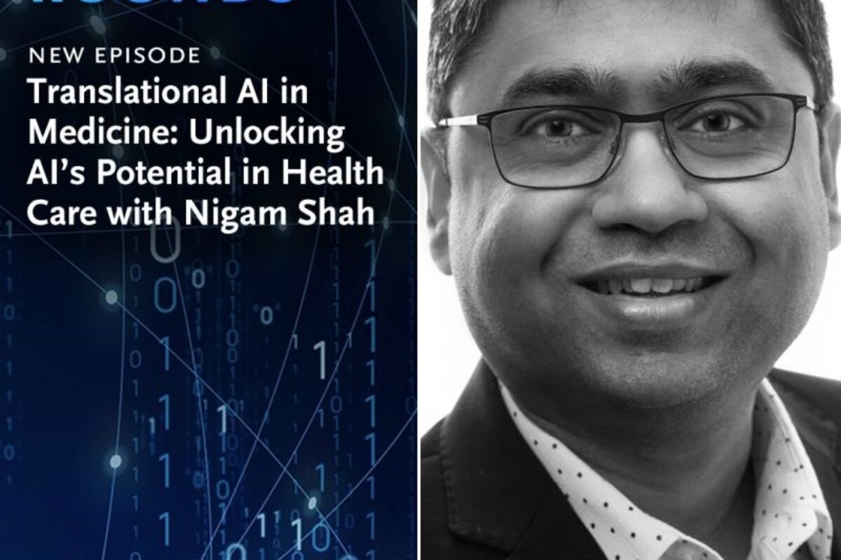
In this episode of the AI Grand Rounds podcast, Dr. Nigam Shah, a distinguished Professor of Medicine at Stanford University and inaugural Chief Data Scientist for Stanford Health Care, shares his journey from training as a doctor in India to becoming a leading figure in biomedical informatics in the United States. He discusses the transformative impact of computational tools in understanding complex biological systems and the pivotal role of hashtagArtificialIntelligence in advancing health care delivery, particularly in improving efficiency and addressing systemic challenges. Dr. Shah emphasizes the importance of real-world integration of AI into clinical settings, advocating for a balanced approach that considers both technological capabilities and the systemic considerations of hashtagAIinMedicine. The conversation with NEJM AI Deputy Editors Arjun Manrai, PhD, and Andrew Beam, PhD, also explores the democratization of medical knowledge, why open-source models are under-researched in medicine, and the crucial role of data quality in training AI systems.

Listen to the full episode: https://nejm.ai/ep18

Congrats

Sohaib Hassan, Elana Simon and Selina Junyi Pi awarded the NSF Graduate Research Fellowship Program

Congratulations to Sohaib Hassan, Elana Simon and Selina Junyi Pi who were all awarded the NSF Graduate Research Fellowship Program (GRFP). The purpose of the program is to help ensure the quality and vitality of the scientific and engineering workforce of the United States. A goal of the program is to broaden participation of the full spectrum of talents in STEM. The five-year fellowship provides three years of financial support inclusive of an annual stipend NSF GRFP.

https://www.research.gov/grfp/AwardeeList.do?method=loadAwardeeList

Congratulations to our DBDS awardees!

Please join us at the next monthly CCSB Seminar, Friday, April 19, from 11 AM to 12 PM. The Stanford Center for Cancer Systems Biology Seminar Series aims to bring together experimental and computational researchers. Speaker: Dr. Sean Bendall, Associate Professor in the Department of Pathology Title: Multi-generational decisions in single cell biology Abstract: Single cell and spatial proteomics, starting with the earliest low parameter fluorescent cytometry and microscopy experiments, helped define the major cell subsets and architecture of human tissues as we understand them today. Now, a novel combination of elemental mass spectrometry with single cell analysis (mass cytometry – CyTOF, Science 2011) and nanometer-scale imaging (multiplexed ionbeam imaging – MIBI, Nature Med. 2014, Cell 2018, Science Adv., 2019) offers routine, simultaneous quantification of > 40 proteomic features without fluorescent agents or interference from spectral overlap and autofluorescence using heavy metal isotopes as reporters. With this, we have reached new levels of understanding in tissue immune organization, combined with novel single-cell visualization and analysis methods. By identifying new cell populations, regulatory relationships, and structural rulesets we have identified numerous clinically predictive features underlying human disease. Location: James H. Clark Center, Room S360, 3rd floor next to the Coffee Shop (our refreshments provider!). Or Online: Zoom link Please contact Corinne Beck if you have any questions and feel free to subscribe to our mailing list here to receive our announcements and updates

The Stanford Center for Cancer Systems Biology Seminar Series: Dr. Sean Bendall, “Multi-generational decisions in single cell biology” 4/19

Please join us at the next monthly CCSB Seminar, Friday, April 19, from 11 AM to 12 PM.
The Stanford Center for Cancer Systems Biology Seminar Series aims to bring together experimental and computational researchers.

SpeakerDr. Sean Bendall, Associate Professor in the Department of Pathology
TitleMulti-generational decisions in single cell biology

Abstract:
Single cell and spatial proteomics, starting with the earliest low parameter fluorescent cytometry and microscopy experiments, helped define the major cell subsets and architecture of human tissues as we understand them today. Now, a novel combination of elemental mass spectrometry with single cell analysis (mass cytometry – CyTOF, Science 2011) and nanometer-scale imaging (multiplexed ionbeam imaging – MIBI, Nature Med. 2014, Cell 2018, Science Adv., 2019) offers routine, simultaneous quantification of > 40 proteomic features without fluorescent agents or interference from spectral overlap and autofluorescence using heavy metal isotopes as reporters. With this, we have reached new levels of understanding in tissue immune organization, combined with novel single-cell visualization and analysis methods. By identifying new cell populations, regulatory relationships, and structural rulesets we have identified numerous clinically predictive features underlying human disease.

Location:  James H. Clark Center, Room S360, 3rd floor next to the Coffee Shop (our refreshments provider!).
Or Online:  Zoom link

Please contact Corinne Beck if you have any questions and feel free to subscribe to our mailing list here to receive our announcements and updates.

Olivier Gevaert and team have developed a biomedical model inspired by DALL-E, we use RNA expression profiles to generate synthetic digital pathology images across several cancer tissues. We show that these synthetic data can be used in combination with real data, cell type distributions are representative of real tissues and synthetic data can be used for self supervised learning. You can try the model here: https://lnkd.in/egWGGDYJ. We also generated 1M images for download: https://lnkd.in/eSsM9ZqA Amazing work by Francisco Carrillo Pérez in the lab, and only possible thanks to the Polaris compute resources and collaboration with Ravi Madduri at Argonne National Laboratory, U.S. Department of Energy (DOE). Full text is available here: https://rdcu.be/dBZJK. https://www.nature.com/articles/s41551-024-01193-8

Olivier Gevaert: “Generation of synthetic whole-slide image tiles of tumours from RNA-sequencing data via cascaded diffusion models” published in Nature Biomedical Engineering

Olivier Gevaert and team have developed a biomedical model inspired by DALL-E, we use RNA expression profiles to generate synthetic digital pathology images across several cancer tissues.
You can try the model here: https://lnkd.in/egWGGDYJ. We also generated 1M images for download: https://lnkd.in/eSsM9ZqA
Amazing work by Francisco Carrillo Pérez in the lab, and only possible thanks to the Polaris compute resources and collaboration with Ravi Madduri at Argonne National LaboratoryU.S. Department of Energy (DOE).
Full text is available here: https://rdcu.be/dBZJK.
https://www.nature.com/articles/s41551-024-01193-8