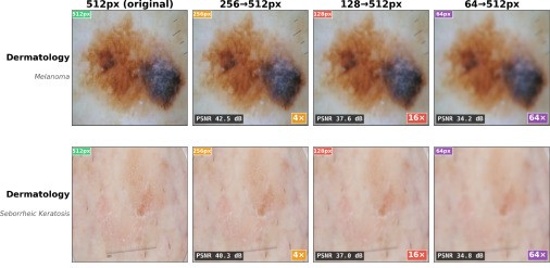
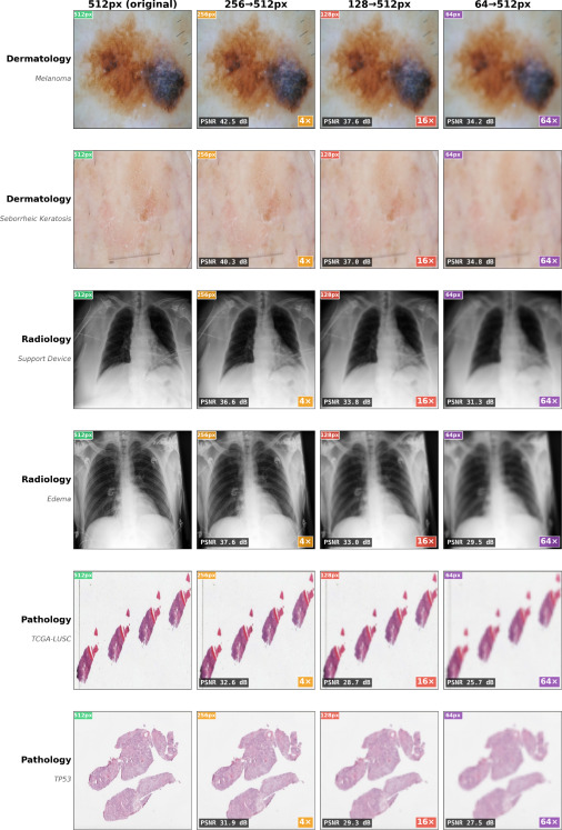
A research team developed Med-REDUCE to explore whether medical AI could work with less image detail and smaller models while maintaining accuracy in distinguishing cancer.
A mole on your skin looks different than normal – could it be skin cancer? If you live in a rural community, getting it checked could mean traveling hours to see a dermatologist. Or not getting it checked at all.
Artificial intelligence (AI) is increasingly being used to help analyze medical images, but these powerful models often need expensive computing equipment. What if a rural clinic could use a handheld skin-imaging device and a simpler AI model to determine whether a mole needs urgent attention?
A new study from researchers at Stanford’s Department of Biomedical Data Science suggests this might someday be possible. In a study published in the journal Medical Image Analysis, scientists led by Vasiliki Bikia, PhD, who collaborated with Sonnet Xu and senior author Roxana Daneshjou, MD, PhD, developed an approach called Med-REDUCE to explore whether medical AI could work with less image detail and smaller models.



Smaller images, smaller models: equally good results
“We were curious: How much of the information in a medical image does AI really need?” said Bikia, then a postdoc in Daneshjou’s lab and now working at Apple. “We kept making the images smaller and less detailed, and were surprised by how well they did. Then we made the models much smaller and got equally good results,” she added.
Medical images contain vast amounts of visual information, but not every pixel is equally important for identifying abnormalities. Some conditions can be recognized from broad shapes and patterns, for example, while others require much finer detail.
Using medical images from dermatology, pathology, and radiology, the researchers tested three AI “vision models” trained to recognize visual patterns. They evaluated how accurately the models could distinguish melanoma from noncancerous skin spots, classify lung and brain tumors, predict certain cancer-related mutations, and identify abnormalities on chest X-rays.
To see how reducing image resolution affected model performance, the researchers progressively reduced the images from 512 × 512 pixels to just 64 × 64 pixels – a 64-fold reduction in the number of pixels. That’s like shrinking an 8-inch-square photograph to just 1-inch square. Most of the fine detail disappears, but the overall shapes and patterns can be recognized.
Even with much less detail, all three original AI models kept most of their accuracy across the medical imaging tasks.
Next, the researchers wanted to see whether they could make the AI models smaller without sacrificing accuracy. They used “knowledge distillation,” a technique in which a larger AI model “teaches” a smaller model what it has learned about recognizing patterns in images.
Across the tasks tested, the smaller models performed as well or better than the larger ones in 39 of 42 comparisons. And in the remaining three tests, the differences in accuracy weren’t significant. Some smaller models performed just as well, despite being only about one-quarter the size of the larger models.
What does all of this mean for patients and their doctors?
Like the example above, lower-resolution medical images could be sent more easily from remote clinics. In the future, portable imaging devices might use smaller AI models that require less computing power. Such advances could help narrow the technological gap between well-resourced academic medical centers and smaller or geographically isolated healthcare facilities.
But reducing image detail has its limits. On chest X-rays, for example, AI could recognize an enlarged heart, but had more trouble detecting a small pocket of air that can cause a lung to collapse. The findings thus highlight the importance of knowing when smaller, simpler AI systems can be used safely – and when more detailed images are truly necessary.
“Even the most powerful AI is of little use if clinics can’t afford it,” said Daneshjou, who is a dermatologist. “One of my longstanding goals is to make medical AI more accessible without sacrificing accuracy or patient safety.”
Med-REDUCE is not yet ready for prime time. This study involved using existing imaging datasets with pretrained AI models. More testing is needed to determine whether such an approach can work reliably in real-world healthcare settings and across different types of patient populations.
But the study points to a future for medical AI in which progress isn’t measured only by how powerful a model is, but by how effectively it can be put to work in more clinics, for more patients.