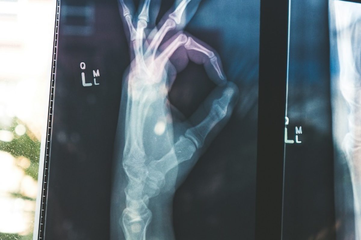
Please join us at the next monthly CCSB Seminar, Friday, April 19, from 11 AM to 12 PM. The Stanford Center for Cancer Systems Biology Seminar Series aims to bring together experimental and computational researchers. Speaker: Dr. Sean Bendall, Associate Professor in the Department of Pathology Title: Multi-generational decisions in single cell biology Abstract: Single cell and spatial proteomics, starting with the earliest low parameter fluorescent cytometry and microscopy experiments, helped define the major cell subsets and architecture of human tissues as we understand them today. Now, a novel combination of elemental mass spectrometry with single cell analysis (mass cytometry – CyTOF, Science 2011) and nanometer-scale imaging (multiplexed ionbeam imaging – MIBI, Nature Med. 2014, Cell 2018, Science Adv., 2019) offers routine, simultaneous quantification of > 40 proteomic features without fluorescent agents or interference from spectral overlap and autofluorescence using heavy metal isotopes as reporters. With this, we have reached new levels of understanding in tissue immune organization, combined with novel single-cell visualization and analysis methods. By identifying new cell populations, regulatory relationships, and structural rulesets we have identified numerous clinically predictive features underlying human disease. Location: James H. Clark Center, Room S360, 3rd floor next to the Coffee Shop (our refreshments provider!). Or Online: Zoom link Please contact Corinne Beck if you have any questions and feel free to subscribe to our mailing list here to receive our announcements and updates

The Stanford Center for Cancer Systems Biology Seminar Series: Dr. Sean Bendall, “Multi-generational decisions in single cell biology” 4/19

Please join us at the next monthly CCSB Seminar, Friday, April 19, from 11 AM to 12 PM.
The Stanford Center for Cancer Systems Biology Seminar Series aims to bring together experimental and computational researchers.

SpeakerDr. Sean Bendall, Associate Professor in the Department of Pathology
TitleMulti-generational decisions in single cell biology

Abstract:
Single cell and spatial proteomics, starting with the earliest low parameter fluorescent cytometry and microscopy experiments, helped define the major cell subsets and architecture of human tissues as we understand them today. Now, a novel combination of elemental mass spectrometry with single cell analysis (mass cytometry – CyTOF, Science 2011) and nanometer-scale imaging (multiplexed ionbeam imaging – MIBI, Nature Med. 2014, Cell 2018, Science Adv., 2019) offers routine, simultaneous quantification of > 40 proteomic features without fluorescent agents or interference from spectral overlap and autofluorescence using heavy metal isotopes as reporters. With this, we have reached new levels of understanding in tissue immune organization, combined with novel single-cell visualization and analysis methods. By identifying new cell populations, regulatory relationships, and structural rulesets we have identified numerous clinically predictive features underlying human disease.

Location:  James H. Clark Center, Room S360, 3rd floor next to the Coffee Shop (our refreshments provider!).
Or Online:  Zoom link

Please contact Corinne Beck if you have any questions and feel free to subscribe to our mailing list here to receive our announcements and updates.

a photo of a sparkler in celebration

Sylvia Plevritis’ five-year anniversary of leading DBDS

Congratulations to Sylvia on leading the Biomedical Data Science department for the past five years. With her strong leadership, direction, and guidance, DBDS is poised to become the leader in the AI/precision healthcare revolution. Sylvia has led the charge for DBDSand has reached milestones in research, discovery, increased grants, and growth with faculty and staff, proving itself to be one of the strongest departments in the School of Medicine. She is tireless, devoted and determined, working incessantly to propel our department to a position of eminence in the biomedical data science field. She is both a visionary and luminary; we could not ask for a more skilled pilot to chart the course for DBDS’ current and future success.

Olivier Gevaert and team have developed a biomedical model inspired by DALL-E, we use RNA expression profiles to generate synthetic digital pathology images across several cancer tissues. We show that these synthetic data can be used in combination with real data, cell type distributions are representative of real tissues and synthetic data can be used for self supervised learning. You can try the model here: https://lnkd.in/egWGGDYJ. We also generated 1M images for download: https://lnkd.in/eSsM9ZqA Amazing work by Francisco Carrillo Pérez in the lab, and only possible thanks to the Polaris compute resources and collaboration with Ravi Madduri at Argonne National Laboratory, U.S. Department of Energy (DOE). Full text is available here: https://rdcu.be/dBZJK. https://www.nature.com/articles/s41551-024-01193-8

Olivier Gevaert: “Generation of synthetic whole-slide image tiles of tumours from RNA-sequencing data via cascaded diffusion models” published in Nature Biomedical Engineering

Olivier Gevaert and team have developed a biomedical model inspired by DALL-E, we use RNA expression profiles to generate synthetic digital pathology images across several cancer tissues.
You can try the model here: https://lnkd.in/egWGGDYJ. We also generated 1M images for download: https://lnkd.in/eSsM9ZqA
Amazing work by Francisco Carrillo Pérez in the lab, and only possible thanks to the Polaris compute resources and collaboration with Ravi Madduri at Argonne National LaboratoryU.S. Department of Energy (DOE).
Full text is available here: https://rdcu.be/dBZJK.
https://www.nature.com/articles/s41551-024-01193-8